Technology Overview

Our team is dedicated to helping people survive cancer. Our passion is focused on enabling the medical community to achieve dramatic improvements in patient survival rates through the early detection of the leading forms of cancer.

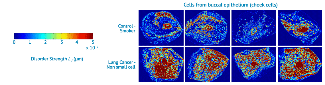
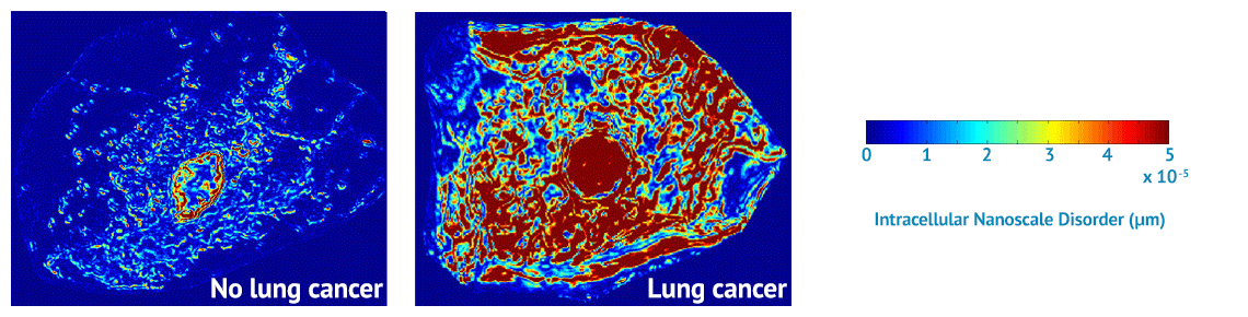
NanoCytomics’ contribution to this effort will be our cancer risk-stratification tests based on our novel, proprietary biophotonics technology platform known as partial wave spectroscopic (PWS) microscopy. Biophotonics deploys optical imaging and sensing technologies to study the structures and functions of cells. Through biophotonics in general and the company’s PWS platform in particular, NanoCytomics can identify cellular abnormalities at the nanoscale level. In so doing, the company anticipates making a profound impact on the ability to stratify cancer risk for an individual patient at a dramatically earlier stage of disease progression, potentially helping physicians to save millions of lives in the process.

NanoCytomics has an exclusive license from Northwestern University for patents and patent applications pertaining to the NanoCytomics technology and methods that were developed in Vadim Backman’s Northwestern Biophotonics Laboratory.

Top

Nanocytology

The American Cancer Society (ACS) defines cytology as “diagnosing diseases by looking at single cells and small clusters of cells…an important part of cancer diagnosis over the past few decades.”  NanoCytomics is building upon the underpinnings of cytology by developing a new optical approach called nanocytology to identify which patients are likely to benefit from gold-standard cancer diagnostic procedures.

Nanocytology is made possible by breakthrough technology (see PWS Platform below) that allows us to see what’s never been seen before. Much of the excitement surrounding this science and engineering is based on the ability to collect cell samples from easily acceptable surrogate sites in the body. For example, nanocytology can detect nanoarchitectural alterations in cells that are obtained by a simple swab of the inside of a person’s cheek. These alterations correlate strongly with the risk of developing lung cancer.

Based on strong clinical data, nanocytology has shown the potential to become a breakthrough risk-stratification platform broadly applicable to almost any organ from which clinically relevant cellular specimens can be obtained (See Field Effect below for why surrogate cells are clinically relevant in cancer).

Top

PWS Platform

We have developed a technology platform within the field of nanocytology that is intended to identify patients that are likely to benefit from gold-standard cancer diagnostic procedures.  Developed by Drs. Vadim Backman and Hariharan Subramanian at the Backman Photonics Laboratory at Northwestern University, partial wave spectroscopic (PWS) microscopy is a breakthrough technology platform that enables quantification of the nanoscale architecture of the cell with unprecedented accuracy.

Cells may appear to be normal to technicians using standard microscopy, but PWS can detect profound changes in the nanoscale architecture of the same cells. PWS measures the disorder strength of the nanoscale organization of the cell, which the NanoCytomics team has determined to be one of the earliest signs of carcinogenesis and a strong marker for the presence of cancer. PWS enables a paradigm shift, in that we don’t need to examine the tumor itself to stratify the cancer risk for an individual patient. (See Field Effect below for why surrogate cells are clinically relevant in cancer).

Our long-term goal is to develop the PWS platform to serve as a highly accurate, low-cost, non-invasive testing to identify which patients are likely to benefit from gold-standard cancer diagnostic procedures.

Top

Field Effect

The company’s technology platform for risk stratification is based on a phenomenon that goes by many names, including “field effect,” “field of injury” and “field carcinogenesis.”  As described by the research community, field effect is the notion that the genetic/environmental milieu that results in carcinogenesis can also be discovered by their impact upon cells in other areas of the body.  The field effect concept is biologically robust and widely used in clinical practice.

Following this principle, published research demonstrate that cells bearing the “fingerprint” of risk (“fertile field”) caused by a malignant tumor are not limited to the tumor site; numerous biomarkers in other areas of the body are altered as well. The challenge has been identifying the presence of these mutations with technology that provides the high levels of sensitivity and specificity (false negatives and false positives, respectively) required to be clinically relevant for large patient populations.

Field carcinogenesis is a general phenomenon reported in multiple cancer types, including lung, colon, breast, ovary, pancreas, prostate. Field carcinogenesis biomarkers fall into one of several categories, such as morphological, micro-architectural,  biochemical, immunohistochemical, cellular, and genomics/epigenetics/proteomics.

However, conventional field effect biomarkers lack the requisite performance for use with large patient populations. Our group has not only confirmed the unprecedented diagnostic accuracy of micro and nano -architectural alterations for the “field effect” but has also devised a clinically compatible methodology for identifying these changes. We have demonstrated PWS-detectable nanoscale alterations in the lung, colon, prostate, ovarian, pancreas and esophagus. Thus, we are confident that PWS will become a new platform for cancer risk stratification that is applicable to almost any organ from which cellular specimen can be obtained by brushing (e.g., cervical, rectal or buccal [cheek] cells) or from secretions (e.g., urine, feces).

Top